Slipped capital femoral epiphysis: Radiographic images may support earlier diagnosis
Slipped capital femoral epiphysis (SCFE) is the most common hip disorder of adolescence, affecting 10.8 per 100,000 children.
Although early intervention leads to better outcomes, SCFE is often not diagnosed in its earliest stages because a lack of clinical suspicion delays diagnosis. Even if a physician suspects the condition, signs of early stage SCFE may not be visible on radiographic images, further complicating the diagnostic process.
A team of researchers at Boston Children’s Hospital, led by orthopedic surgeon Eduardo Novais, MD, have identified a radiographic sign that could help clinicians identify the condition earlier.

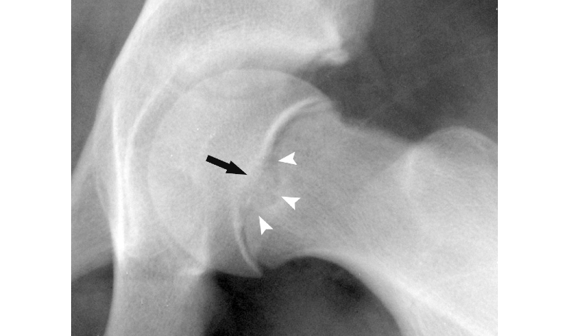
Figure 1: Peritubercle lucency on a radiograph
Understanding the mechanisms of SCFE
The investigation grew out of a series of studies elucidating the mechanisms of SCFE. Whereas the condition has traditionally been described as a slip of the capital femoral epiphysis over the metaphysis, recent studies have suggested that the displacement is actually a matter of rotation, with the epiphyseal tubercle serving as a fulcrum for rotation. If so, standard methods, such as the Klein method or Southwick angle that look for signs of slippage, would not recognize the type of subtle displacement that takes place during early stages of SCFE.
The team at Boston Children’s theorized that if the epiphyseal tubercle stabilizes the epiphysis, the abnormal mechanical environment associated with SCFE could lead to a concentration of stress around the tubercle. Such stress could cause changes in the area that could be identified on radiographs. In a recent study, the team evaluated the presence of peritubercle lucency (Figure 1) as an indicator of micro-instability around the epiphyseal tubercle and, more importantly, early stage SCFE.

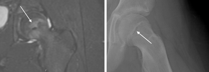
Figure 2: MRI (left) showing peritubercle edema suggestive of SCFE compared to a radiographic image of the same hip with peritubercle lucency
Radiographic images compared to MRI and a new classification of SCFE
To test its theory, the team enlisted five independent observers to blindly assess the radiographs of 71 patients for the presence or absence of peritubercle lucency. All patients had previously undergone MRI for an evaluation of pre-slip or minimally displaced SCFE. Sixty percent (43) had confirmed SCFE.
Comparing radiographic images with indications of SCFE on MRI (Figure 2), the team found that the presence of the peritubercle lucency sign on radiographs is accurate, with high sensitivity and specificity for the diagnosis of a pre-slip condition or minimally displaced SCFE.
The team also proposed a new classification for SCFE based on the relationship of the epiphyseal tubercle and the metaphyseal fossa (Figure 3).
Implications for diagnosis and treatment
This research is ongoing. In the future, the results could help orthopedists diagnose SCFE earlier and initiate clinical intervention before the condition has a chance to progress. Further, a more complete understanding of the mechanisms of SCFE could have future implications for clinical and surgical treatments.

Figure 3: Proposed new SCFE classification based on the relationship of the tubercle and its metaphyseal socket
Articles referenced
Maranho DA, Bixby SD, Miller PE, Hosseinzadeh S, George M, Kim YJ, Novais EN. What Is the Accuracy and Reliability of the Peritubercle Lucency Sign on Radiographs for Early Diagnosis of Slipped Capital Femoral Epiphysis Compared With MRI as the Gold Standard? Clin Orthop Relat Res. 2020 Jan 21.
Maranho DA, Bixby S, Miller PE, Novais EN. A novel classification system for slipped capital femoral epiphysis based on the radiographic relationship of the epiphyseal tubercle and the metaphyseal socket. J Bone Joint Surg Open Access. 2019;4: e0033.
Maranho DA, Miller PE, Novais EN. The Peritubercle Lucency Sign is a Common and Early Radiographic Finding in Slipped Capital Femoral Epiphysis. J Pediatr Orthop. 2018 Aug; 38(7):e371-e376.
Related articles
Novais EN, Maranho DA, Vairagade A, Kim YJ, Kiapour A. Smaller Epiphyseal Tubercle and Larger Peripheral Cupping in Slipped Capital Femoral Epiphysis Compared with Healthy Hips: A 3-Dimensional Computed Tomography Study. J Bone Joint Surg Am. 2020 Jan 02; 102(1):29-36.
Kiapour AM, Kiapour A, Maranho DA, Kim YJ, Novais EN. Relative contribution of epiphyseal tubercle and peripheral cupping to capital femoral epiphysis stability during daily activities. J Orthop Res. 2019 07; 37(7):1571-1579.
Novais EN, Maranho DA, Kim YJ, Kiapour A. Age- and Sex-Specific Morphologic Variations of Capital Femoral Epiphysis Growth in Children and Adolescents Without Hip Disorders. Orthop J Sports Med. 2018 Jun; 6(6):2325967118781579.
