Our team is always looking for ways to improve brain surgery and make the experience easier for children and their families. Our breakthroughs — taking place in our clinics, as well as in our basic science laboratories — play a critical role in your child’s health. We are continually developing and refining minimally invasive techniques as alternatives to traditional brain and spinal cord surgeries.
Our team is also actively involved in research projects to improve understanding of why brain diseases occur, and to help develop better methods of early detection to ensure that these conditions have as little impact as possible on a child’s developing brain.
A few of our recent innovations include:
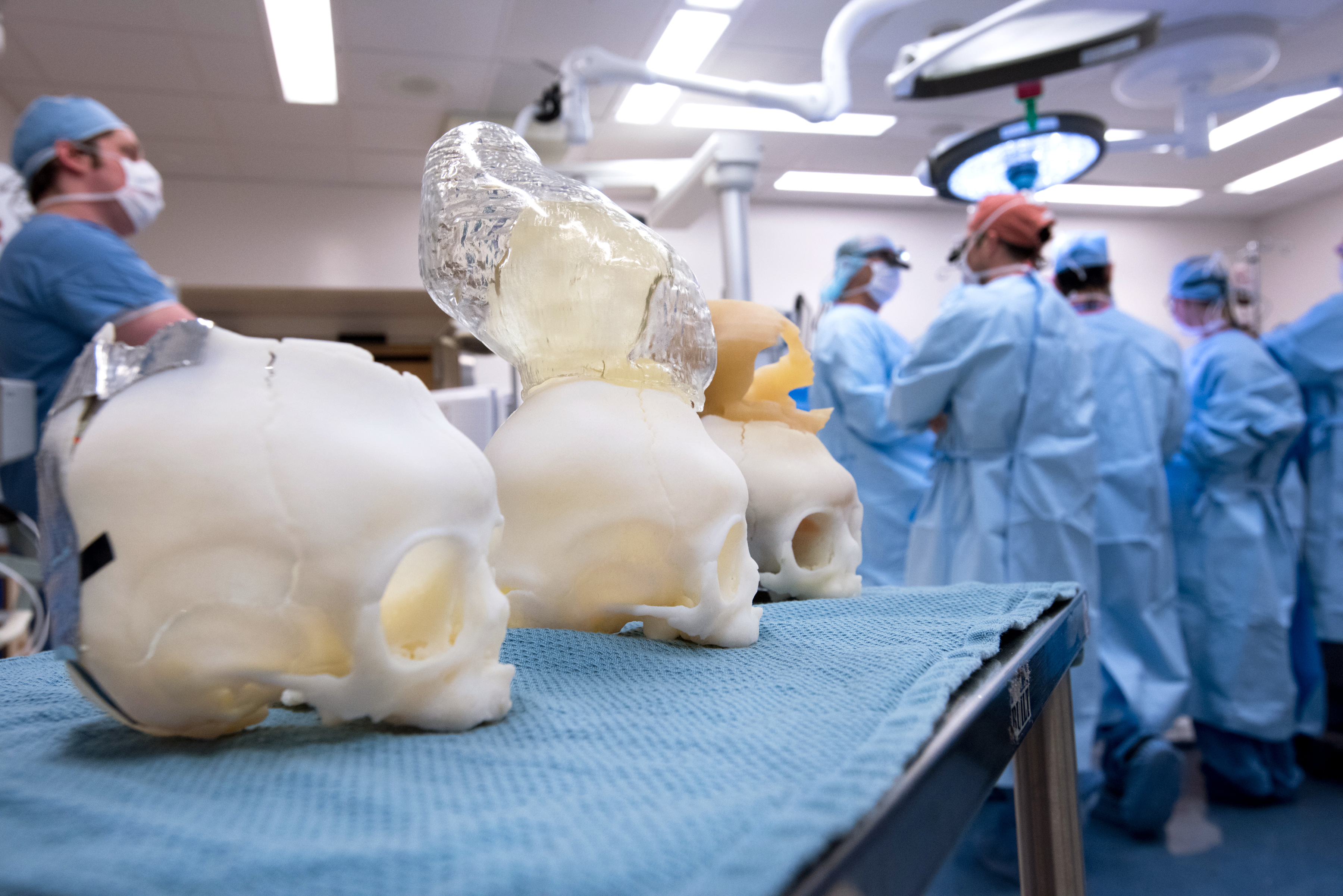
3-D printing for surgical planning
The neurosurgery team was an early adopter of three-dimensional printing technology, collaborating closely with Boston Children’s Immersive Design Systems. Engineers can quickly produce life-sized models of patients’ brains, which allows surgeons to more precisely plan surgery and to rehearse different approaches. The 3-D models provide details not normally visible to surgeons before surgery.
ETV/CPC for hydrocephalus
In 2000, one of the neurosurgeons on our team, Dr. Benjamin Warf, pioneered a treatment for hydrocephalus in infants as an alternative to shunting. The treatment, called ETV/CPC, combines two procedures: endoscopic third ventriculostomy (ETV) and choroid plexus cauterization procedure (CPC). Published research has demonstrated that ETV/CPC continues to provide good outcomes for babies with hydrocephalus.
Pial synangiosis
Pial synangiosis is a type of surgery for moyamoya that was developed at Boston Children’s in 1985. Since that time, it has become the mainstay of treatment for moyamoya. Most children who have pial synangiosis are released from the hospital within a few days, and usually need only regular exams and monitoring as follow-up. Long term outcomes demonstrate patients can lead productive, independent lives following surgery.
Laser therapy for deep brain lesions
Boston Children’s is one of a handful of centers offering a new, minimally invasive laser therapy or laser ablation surgery to remove tumors or diseased brain tissue that is too deep inside the brain to safely access with usual neurosurgical methods. This procedure is an excellent option for children with deep brain tumors or seizures caused by abnormalities deep within the brain, who have not been helped by medication and for whom surgery would normally be considered unsafe.
SEEG
Boston Children’s is a leading destination for pediatric stereoelectroencephalography (SEEG) in the U.S. SEEG is a procedure in which electrodes are fed on tiny wires through paths drilled into the skull. These electrodes can reach deeper into a child’s brain — and can present a better opportunity for clinicians to determine the source of seizure activity. SEEG is less invasive and better tolerated in kids. When coupled with robotic guidance, it reduces time in the operating room and allows surgeons to implant electrodes in places not typically possible.
