Current Research | Overview
Project One
Infantile hemangioma is a vascular tumor that can grow rapidly, causing organ damage, disfigurement and morbidity. My lab has focused on the cellular mechanisms that drive this uncontrolled vascular growth. We isolated increasingly less differentiated cells from hemangioma tissue, which led us to the identification of a multi-potent stem cell that can recapitulate hemangioma in immune-deficient mice. The hemangioma stem cells differentiate into endothelial cells, pericytes and adipocytes, thereby forming the major cell types in this common childhood tumor.
We have three major goals in this project. The first is to understand the mechanisms that control the differentiation of the hemangioma stem cells into endothelial cells, pericytes and adipocytes. The second is to determine if there are somatic mutations that cause or contribute to infantile hemangiom. The third is to understand how endothelial-pericyte interactions in hemangioma contribute to the spontaneous involution of the tumor.
Project Two
Vascular malformations are defects in the architecture and function of arteries, veins, capillaries and lymphatic vessels. The malformations can be familial or sporadic, and are often associated with tissue overgrowth, deformity and infections. We focus on capillary malformations (CM), which consist of excessive and abnormal capillary/venule-like vessels. CMs are often on the face and are referred to in lay terms as port-wine stains. Sturge-Weber syndrome (SWS) is a rare neurological disorder in which excessive abnormal vessels akin to CM are found on the surface of the brain, and are thought to contribute to the neurologic deficits in SWS.
A somatic, activating mutation in GNAQ (p.R183Q) was discovered in patients with SWS and CM, linking these two disorders. We fractionated CM lesions and SWS brain lesions into specific cell populations that were then genotyped for the GNAQ (p.R183Q) mutation. Our studies show that the GNAQ (p.R183Q) mutation is enriched in the endothelial cells from skin and brain CMs. This identification the cellular context in which the GNAQ mutation resides provides essential information needed to decipher the mechanisms by which CMs form, how to prevent CM and how to regress CM.
We have three major goals in this project. The first is to understand how the GNAQ mutation in endothelial cells leads to the excessive and abnormal blood vessels. The second is to determine if GNAQ mutant endothelial cells exert paracrine effects on surrounding cells that are deleterious and the third is to create models that reflect the impacts of mutant GNAQ so that we can screen for drugs to treat SWS.
Project Three
We also study endothelial plasticity in cardiac valves. We showed that valve endothelial cells from aortic, pulmonic and mitral valves can recapitulate processes that occur during valve development such as endothelial to mesenchymal transition (EndMT). We propose that the EndMT-competent cells along the valve endothelium are valve progenitor cells and that their job is replenish the valve interstitial cells on an as-needed basis throughout adult life and possibly during valve disease.
Together with our collaborators Drs. Levine and Aikawa, we study EndMT in the mitral valve after myocardial infarction (MI) in sheep to determine the extent to which EndMT might be part of the adaptative mechanism to limit mitral regurgitation after MI. Our recent paper (Circ Res 2016) shows that the protein tyrosine phosphatase CD45 is expressed in vivo in mitral valve endothelial cells after MI. In vitro experiments demonstrate that CD45 phosphatase is needed for mitral valve endothelial cells to undergo EndMT. This novel discovery indicates that CD45 is an important regulator of EndMT.
We have three major goals in this project. The first is to determine the extent to which valve endothelial cells contribute, directly or indirectly, to valve fibrosis. The second is to identify up and downstream targets of CD45 in valve endothelial cells. Our third goal is to determine if CD45 participates in other settings of pathological EndMT.
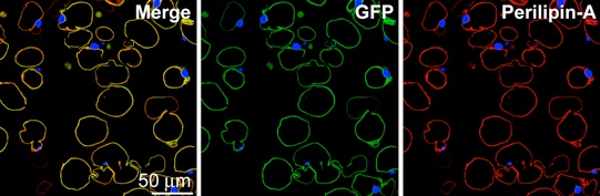 Hemangioma-derived stem cells form adipocytes two months after implantation in vivo (from Khan, Boscolo et. al., J. Clin. Invest. 2008.)