Clubfoot | Diagnosis & Treatments
How is clubfoot diagnosed?
Most of the time, a baby’s clubfoot is diagnosed during a prenatal ultrasound before they are born. About 10 percent of clubfeet can be diagnosed as early as 13 weeks into pregnancy. By 24 weeks, about 80 percent of clubfeet can be diagnosed, and this number steadily increases until birth.
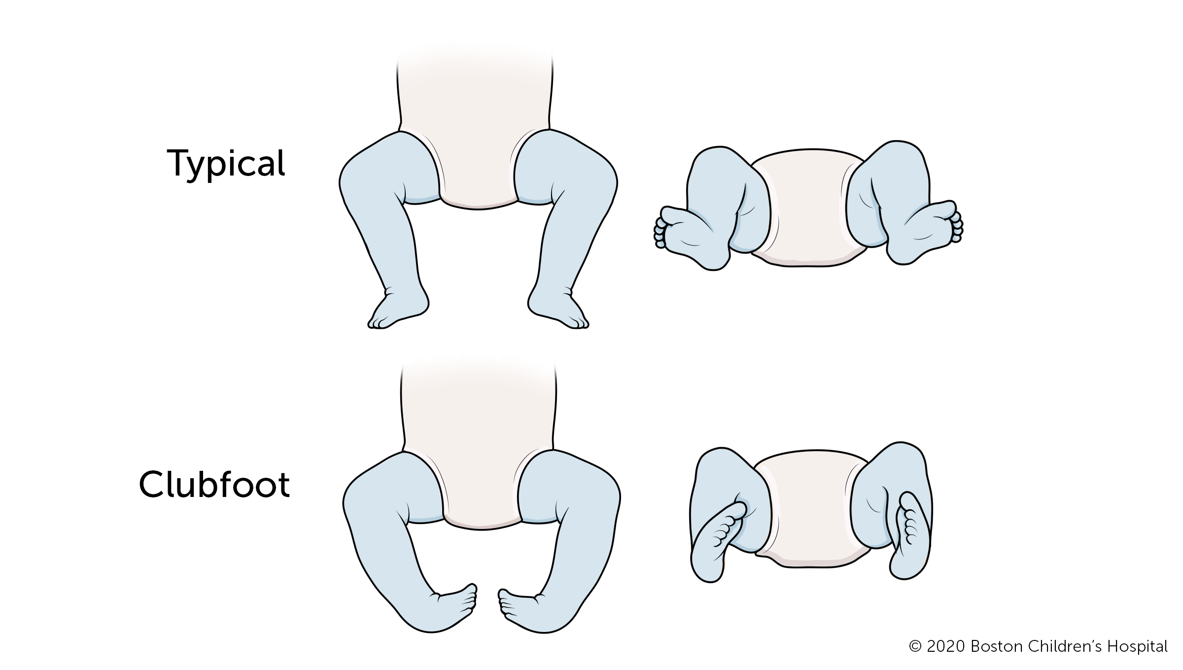
If a child is not diagnosed before birth, clubfoot can be seen and diagnosed as soon as they are born. A physical exam is generally all that is necessary to establish a diagnosis. In rare cases other tests may be used, including:
- x-ray
- computerized tomography scan (CT or CAT scan)

When your baby has clubfoot: Advice for expecting parents
“Our goal when we meet with parents is to reassure them that clubfoot is treatable.”
How is clubfoot treated?
The goal of clubfoot treatment is to correct the position of the foot so that the bones, tendons, and muscles of the foot can grow more normally. Ideally, treatment begins within one month of a child’s birth, when their feet and ankles are at the earliest possible stage of development.
Ponseti method
The Ponseti method is the most common and effective clubfoot treatment. This treatment uses a series of casts and braces to rotate the baby’s foot into a corrected position. The foot is rotated externally until it is turned out 60-70 degrees. Treatment usually begins sometime between birth and 4 weeks of age and involves two stages: treatment and bracing.
Treatment stage
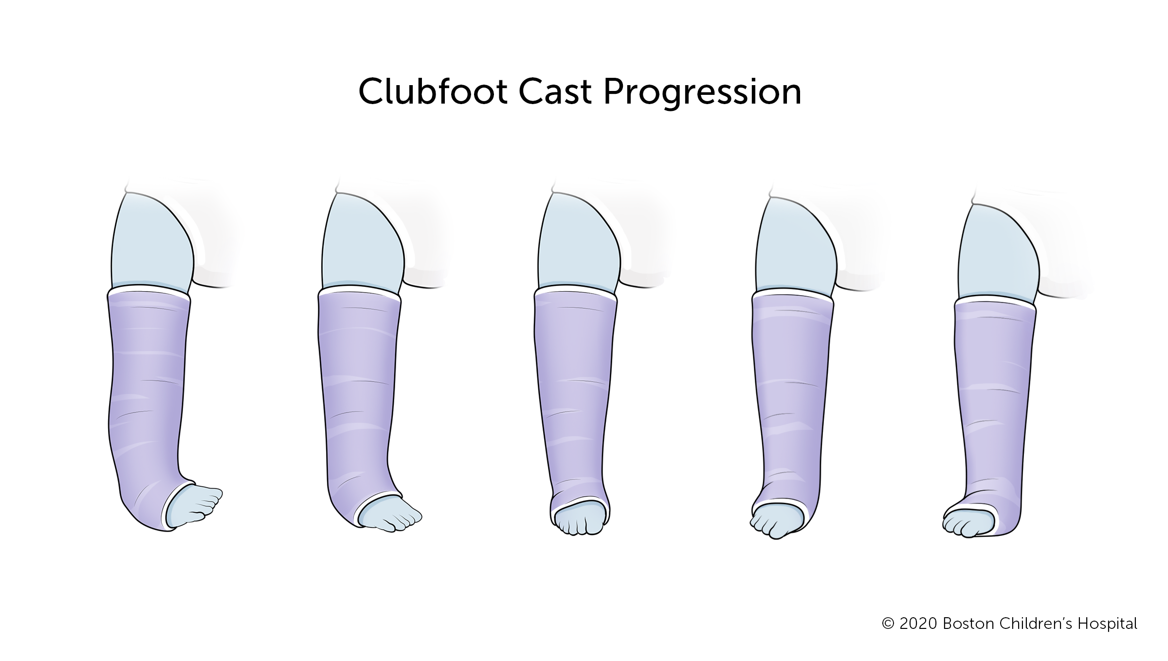
During the treatment stage, your child’s doctor will slowly reposition your child’s foot using a series of casts. This stage involves two to three months of stretching and repositioning the foot.
- The doctor will stretch and reposition your child’s foot, then place their foot, ankle, and leg in a cast to hold the foot in the new position.
- The cast will be removed after about a week, and the doctor will once again reposition your child’s foot. A new cast will hold the foot in its new position.
- This process will be repeated every week until your child’s foot is moved from its incorrect inward-facing position to the correct outward position. Typically, it takes five to eight readjustments and cast changes to move the foot into a correct position.
- When the foot is in its improved outward position, most children need minor surgery (tenotomy) to lengthen their Achilles tendon. This is the cord that attaches the calf muscle to the heel. About 95 percent of babies need this surgery, which is usually performed under local anesthesia.

See tips for cast care and maintenance.
Clubfoot bracing stage
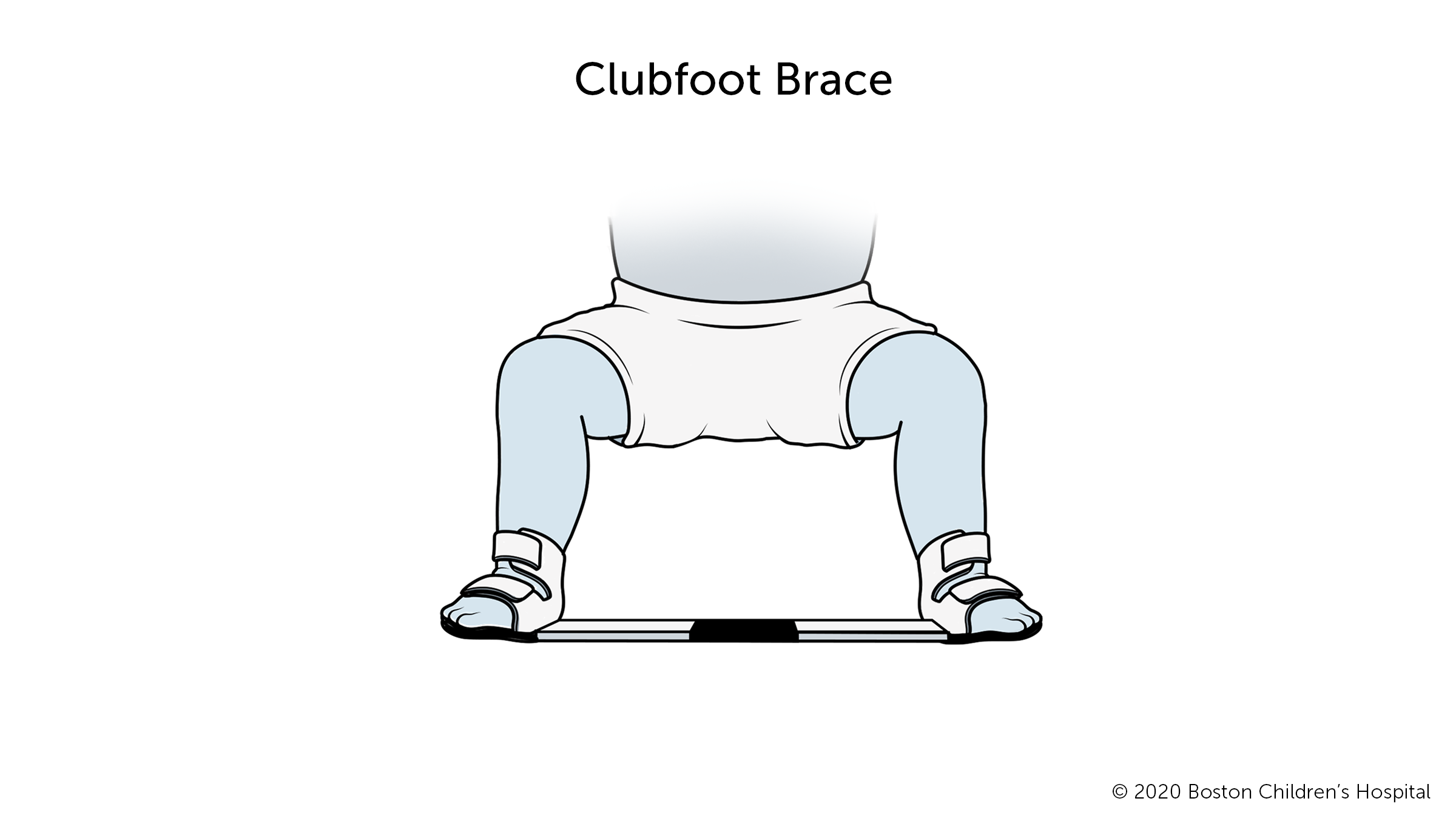
Clubfoot bracing lasts for several years and is crucially important to your child’s long-term mobility. The brace maintains your child’s foot in a corrected position.

- From the end of the treatment stage until your child is 3 to 6 months old, they will wear the brace about 22 hours a day.
- After this initial period, your child’s doctor will probably say it’s OK for your child to wear the brace at night and at nap time, about 15 or 16 hours a day.
- When your baby is ready to learn how to crawl, and then walk, run, and play, they can do so with the brace off.
- You will need to follow the bracing program strictly until your child is 4 years old. Despite the inconvenience, this is the best way to prevent your child’s foot from turning inward again and needing further medical intervention.
What is the long-term outlook for babies with clubfoot?
With early treatment and bracing, almost all babies with clubfoot grow up to have normally functioning feet. They can run, play, and wear normal shoes. If only one foot is affected, it will most likely be smaller and somewhat less mobile than the other foot. Your child may require two different shoe sizes. The affected leg may be slightly smaller and the calf may be less muscular than their other leg.
While clubfoot responds well to treatment, it does not get better on its own. If left untreated, clubfoot will become worse with age and make it hard for your child to walk. Therefore, early treatment and following the bracing program closely are very important.
How we care for clubfoot at Boston Children’s Hospital
The Lower Extremity Program at Boston Children's Hospital takes a conservative, non-surgical approach to clubfoot whenever possible, and we have excellent success rates. In the rare case that a newborn needs surgery, we work with the Department of Anesthesiology, Critical Care and Pain Medicine to avoid the use of general anesthesia whenever we can.
We offer an especially strong and supportive bracing program. We also work with the Fetal Care and Surgery Center, the hospital’s prenatal counseling program, to help parents anticipate and plan for their baby’s care after birth.


