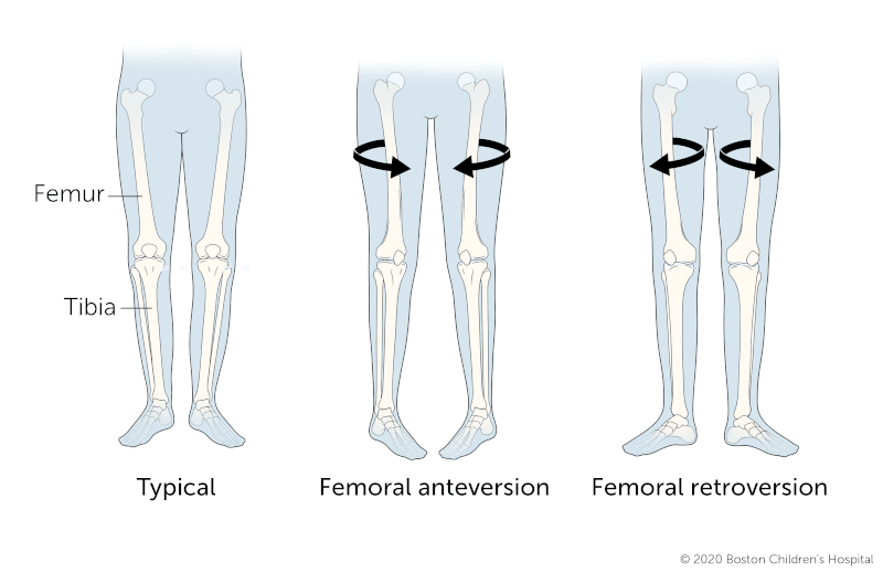
Femoral Anteversion | Diagnosis & Treatments
How is femoral anteversion diagnosed?
Your child’s doctor will conduct a physical exam to diagnose femoral anteversion. As part of the exam, the doctor will take your child’s complete prenatal, birth, and family medical history.
The physical exam also includes measurements to determine the degree of your child’s in-toeing. These measurements can be obtained easily just by placing ink or chalk on the bottom of your child's feet and having them walk on paper to leave an impression.
The doctor may use diagnostic tests to get detailed images of your child’s thigh bone and hip joint, including:
- Computed tomography (CT or CAT) scan uses x-ray equipment and powerful computers to create detailed, cross-sectional images of your child's body. A CT scan is the main imaging test used to confirm a diagnosis of femoral anteversion. It is faster than an MRI and more detailed than x-rays.
- MRI (magnetic resonance imaging) uses magnets, radio frequencies, and a computer to produce detailed images of organs and structures within your child’s body.
- X-rays use invisible electromagnetic energy beams to produce images of internal tissues, bones, and organs.
How is femoral anteversion treated?
Doctors treat most children who have femoral anteversion with close observation over the course of several years. For most children, the twisting of the thigh bone usually corrects by itself with time. Most children achieve normal or near-normal walking patterns by the time they are 8 to 10 years old. Others achieve normal walking patterns by the time they reach their teen years.
Braces, special shoes, and exercises don't usually speed up or help the body's own mechanism for self-correcting femoral anteversion. However, your child’s doctor may recommend one of these treatments if your child’s feet point inward severely or if the condition is not getting better over time.
What are the surgical options for femoral anteversion?
In a very few cases, the twisting-in may be severe and may not self-correct by the time a child is age 8 or 9. For children with severe, unresolved femoral anteversion at that age, doctors may perform surgery to reposition the femur at a more normal angle.
During the procedure (called a femoral derotation osteotomy) the surgeon cuts the femur, rotates the ball of the femur in the hip socket to a normal position, and reattaches the bone.
What is the recovery process from femoral anteversion surgery?
After surgery, your child will probably stay in the hospital for a couple of days and be given pain medication. When your child goes home, they'll need to limit weight-bearing activities, and might use crutches or a walker for a few weeks. Physical therapy will help restore your child’s muscle strength. They'll probably be able to resume full activities, including sports, after three or four months.
What is the long-term outlook for children with femoral anteversion?
Femoral anteversion is self-correcting in up to 99 percent of cases, and the long-term outlook is very positive for most children with the condition. Femoral anteversion doesn't typically lead to arthritis or any other future health problems.
The outlook is also excellent for children with a severe form of the condition who need surgery. The surgery itself is quite safe. In addition, children's bones usually heal faster and more reliably than adults.


